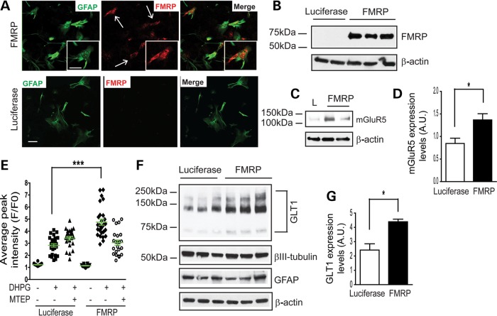
Figure 8.
Re-expression of FMRP in fmr1−/− astrocytes rescues astroglial mGluR5 expression/function and neuron-dependent GLT1 expression. Re-expression of FMRP in cultured fmr1−/− astrocytes is evidenced by FMRP immunostaining (A) and immunoblot (B). Luciferase (L) cDNA was used as a negative control. A magnified view was shown in the insert image. Scale bar: 50 μm; (C) a representative mGluR5 immunoblot from FMRP re-expressed fmr1−/− astrocytes; (D) quantification of the mGluR5 expression in FMRP re-expressed fmr1−/− astrocytes. n = 4. *P < 0.05 from Student's t-test; (E) DHPG-induced Ca2+ responses in FMRP re-expressed astrocytes n = 24–27 astrocytes/group ***P < 0.001 from one-way ANOVA with the Bonferroni post-test; (F) Neuron-dependent GLT1 expression in FMRP re-expressed astrocyte and wild-type neuron co-cultures; Wild-type neurons were plated 3 days following transfection. Luciferase cDNA transfection serves as a control. (G) Quantitative analysis of GLT1 protein expression levels following FMRP re-expression in co-cultures. n = 6–8 independent transfections/group, *P < 0.05 from Student's t-test.

