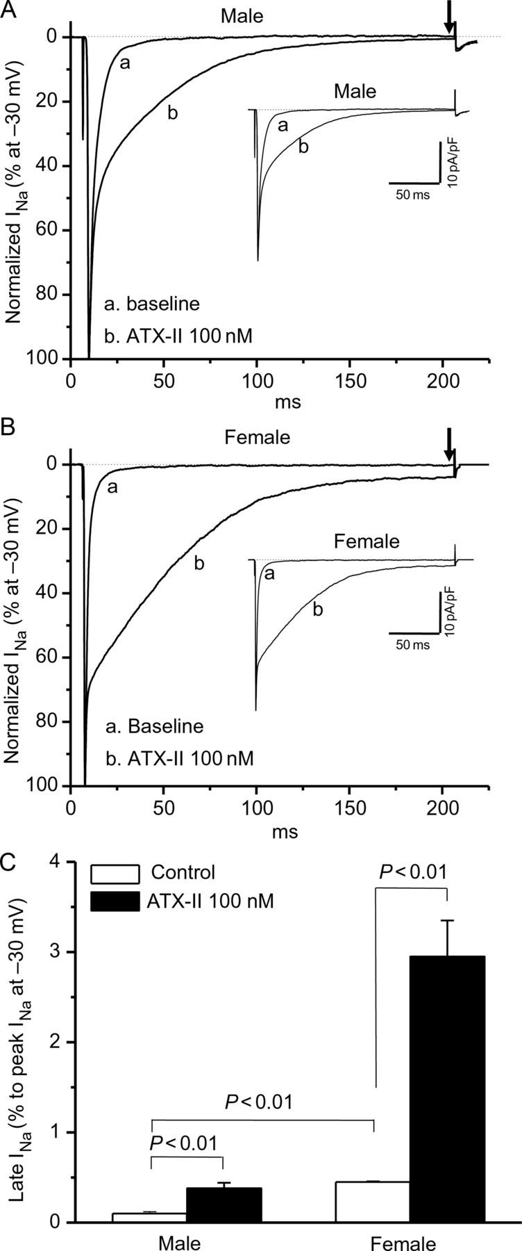
Figure 3.

Late sodium current (INa-L) in male and female ventricular myocytes. Late sodium current in male (A) or female (B) myocytes with or without the addition of ATX-II. Female myocytes have a significantly larger late current at baseline that is further increased by ATX-II treatment. Late current was measured as a percentage of peak current before the ending of 200 ms pulsing after peak INa (indicated by arrows), while non-normalized raw current traces are shown in insets. Late current data (n = 6 each) are summarized in (C).
