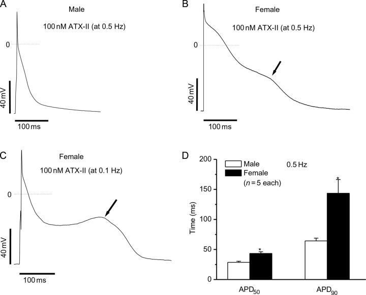
Figure 5.
Comparisons of ventricular action potentials from male and female mice at slow stimulation frequencies in the presence of ATX-II. Action potentials were recorded from isolated cardiomyocytes in male (A) and (B) female mice. With slowed frequency (0.5 Hz), alterations in the trajectory of late repolarization (arrows) were not observed in cells from the male mice (0/5), but were recorded in cells from female mice (3/5). (C) At an even slower rate, 0.1 Hz, a more prominent discontinuity of late repolarization was seen in female myocytes (3/5). At slow stimulation rate, ATX-II-induced APD prolongations were greater in female mice, as summarized in (D) (*P < 0.01).

