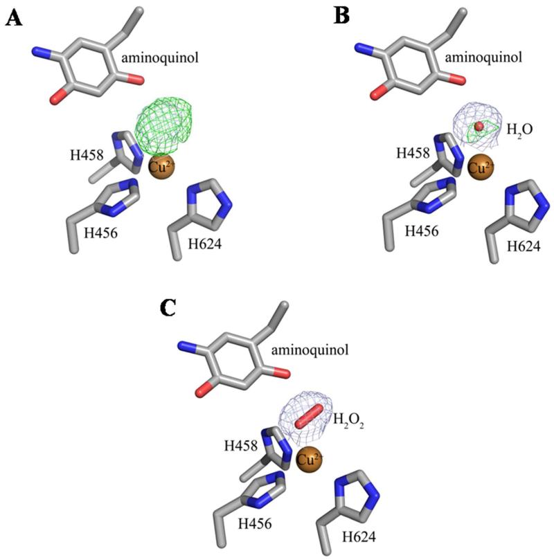
Figure 2.
Structures of the active site of chain E of the ethylamine-HPAO-1 complex with (A) no model, (B) water (Wa), or (C) hydrogen peroxide (H2O2) modeled as the axial copper ligand. Residues in all panels and the peroxide in panel (C) are drawn in stick and colored by atom type (carbon, grey). Copper ions in all panels are drawn as gold spheres and the water molecule in panel (B) is drawn as a small red sphere. The 2Fo-Fc electron density maps after refinement against the corresponding species are drawn as a blue mesh and contoured at 1σ. The Fo-Fc electron density maps after refinement are drawn as a green mesh and contoured at +3.5σ.

