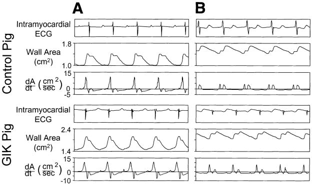
Fig. 6.

Typical recordings of regional wall area and the first derivative of wall area with respect to time (+dA/dt). Top: recordings from ischemic zone of a pig from the control group. Bottom: recordings from ischemic zone of a pig from the GIK group. For both pigs, recordings are shown under baseline conditions (A) and after 90 min reperfusion after 90 min ischemia (B). Under baseline conditions, both pigs exhibit rapid diastolic wall expansion and no evidence of early systolic wall expansion. After reperfusion, the pig from the control group exhibits impaired early diastolic wall expansion (reduced +dA/dtmax); early systolic wall expansion is also evident. In comparsion, the GIK-treated pig exhibits greater recovery of +dA/dtmax and less prominent early systolic wall expansion. Note that absolute values of wall area and dA/dt vary between pigs because of differences in distance between pairs of ultrasonic crystals at implantation.
