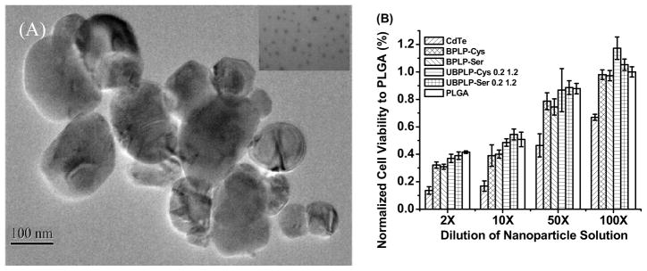
Figure 8.

Morphology and cytotoxicity study of UBPLPs nanoparticles. (A) TEM images of UBPLP-Ser 1.2 nanoparticles. Inset is an image captured under higher magnification showing evenly dispersion of nanoparticles; (B) Evaluation of cytotoxicity of BPLPs and UBPLPs nanoparticle solutions at different dilution. PLGA nanoparticles were used as a control.
