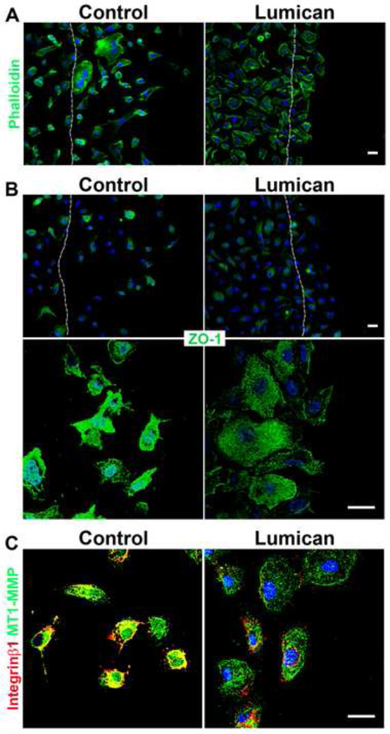
Figure 6.
Role of lumican on the expression and localization of actin filaments, ZO-1, integrin β1 and MT1-MMP in prostate cancer cells. Coverslips placed in 24 well polystyrene culture dishes were incubated in a solution of lumican (10 µg/mL) and untreated culture dishes were used as controls. After culture dish treatment, 2 × 10⁴ prostate cancer cells, PC3, were seeded in RPMI containing L-glutamine/penicillin/streptomycin and 10% FBS, and incubated for 24 h at 37°C in a 5% CO2 humidified environment. A scratch was performed in the cells on the culture dish using a 100 µL pipette tip. Dashed line represents the original wound edge. Cells were washed three times with EBSS, maintained for a further 4 h (37°C, 5% CO2) and fixed for immunocytochemistry. The cells were labeled with phalloidin (A) and anti-ZO-1 (B) and the nuclei were visualized with DAPI. Images were captured using a Zeiss LSM510 scanning Confocal inverted microscope and Zeiss Observer Z1 microscope coupled with an ApoTome.

