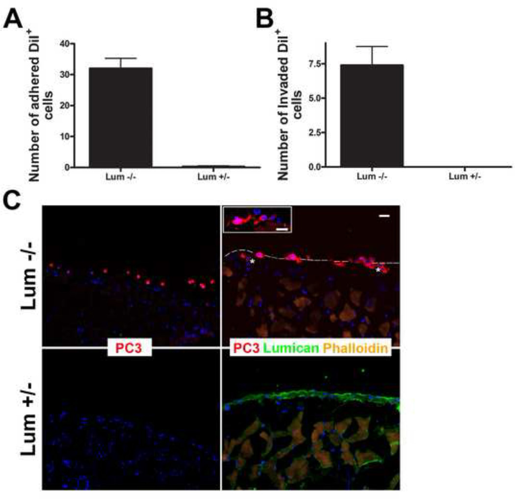
Figure 8.
Role of lumican on the capacity of prostate cancer cells to invade adjacent tissue. Prostate cancer cells (PC3) previously labeled with DiI (red) were seeded upon peritoneal tissue isolated from either lumican KO mice or littermate heterozygous controls and left to invade for 24 h. The peritoneum was washed and fixed and the invaded cells were visualized under a fluorescent microscope. The total number of DiI positive cells which had penetrated either lumican KO or littermate control tissue were counted and represented in a graph (A). PC3 cells adhered to and invaded the lumican KO peritoneal tissue, whereas few cells adhered to the heterozygous tissue and no cells fully invaded the heterozygous tissue (B). PC3 cellular projections (asterisk) protrude into the lumican KO peritoneal tissue.

