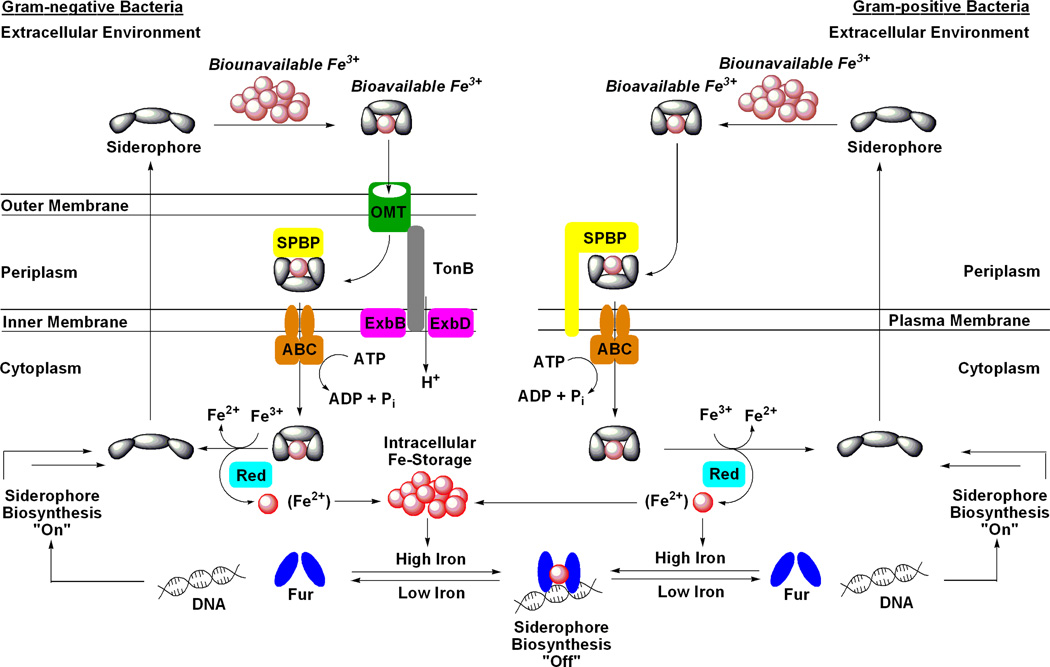
Figure 1.
Generic schematic of siderophore-mediated iron uptake and genetic regulation in Gram-negative and Gram-positive bacteria. Iron metabolism in bacteria is under genetic control by the ferric uptake regulator (Fur) transcription factor protein. During times of iron abundance, the Fur protein complexes Fe(II) and adopts a conformation that binds to a region of DNA known as a ‘Fur box’ which represses the expression of siderophore-related proteins, including biosynthetic enzymes. During times of iron deficiency, the apo-Fur protein dissociates from the ‘Fur box’ allowing expression of siderophore-related genes. The siderophore system is now fully functional and begins with siderophore biosynthesis and efflux to the extracellular environment. For Gram-negative bacteria, after the siderophore chelates Fe(III) to form a soluble complex it is transported into the periplasm by a high affinity outer membrane transport protein (OMT; formerly called OMR). This transport is driven by the proton motive force with energy transfer mediated by the membrane-spanning TonB-ExbB/D protein complex. The Fe(III)-loaded siderophore is then bound by a siderophore periplasmic binding protein (SPBP) and trafficked to an ATP-binding cassette transporter (ABC) that moves the siderophore-Fe(III) complex into the cytoplasm. The iron nutrient is then removed from the siderophore, typically via reduction of Fe(III) to Fe(II) by an iron reductase enzyme (Red), and is distributed to parts of the cell in need or stored in bacterioferritin. In most cases, the siderophore is then recycled for further use. Gram-positive bacteria lack the outer membrane found in Gram-negative bacteria and therefore also lack the OMTs and TonB complex. Instead, extracellular siderophore-Fe(III) complexes are recognized and transported by membrane-anchored SPBPs. The remaining steps of the siderophore pathway in Gram-positive bacteria are analogous to those described for Gramnegative bacteria. This figure was inspired by a variety of siderophore transport diagrams reported in the literature which also describe these pathways in great detail.(13–15)

