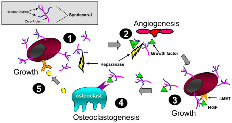
Figure 1. Model showing syndecan-1 expression and function in the myeloma microenvironment.
(1) Myeloma cells shed syndecan-1 that accumulates adjacent to cells and within bone marrow stroma (2), where it traps and concentrates growth and chemotactic factors and stimulates angiogenesis. Heparanase fuels the process by enhancing syndecan-1 shedding and releasing matrix-bound growth factors. This growth factor enriched microenvironment supports further seeding of myeloma cells in the stroma (3) that proliferate in response to signaling via the HGF/syndecan-1/cMET complex and others. These proliferating myeloma cells release factors stimulating osteoclastogenesis (4). Once activated, osteoclasts secrete additional unknown factors that further stimulate myeloma growth (5). This model depicts only a fraction of interactions between syndecan-1 and heparan sulfate in the myeloma microenvironment, it is likely that many others exist. The gray box illustrates syndecan-1 structure.

