Fig. 5.
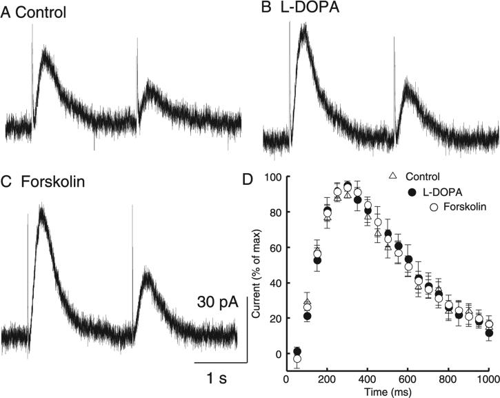
Increasing the amplitude of the IPSC did not change paired-pulse depression or IPSC kinetics. A pair of single pulses 2 s apart (A) always yielded paired-pulse depression that (B) did not change when vesicular content was increased with L-DOPA (10 μm) or (C) release probability was increased with forskolin (10 μm). These manipulations did yield larger amplitude IPSCs (control, 26.2 ± 3.0 pA; L-DOPA, 36.6 ± 4.7 pA; forskolin, 64.2 ± 14.3 pA; n = 9–11). (D) Normalizing the traces to their maxima revealed that the increase in size was not accompanied by a change in time course of the IPSC (n = 6–11).

