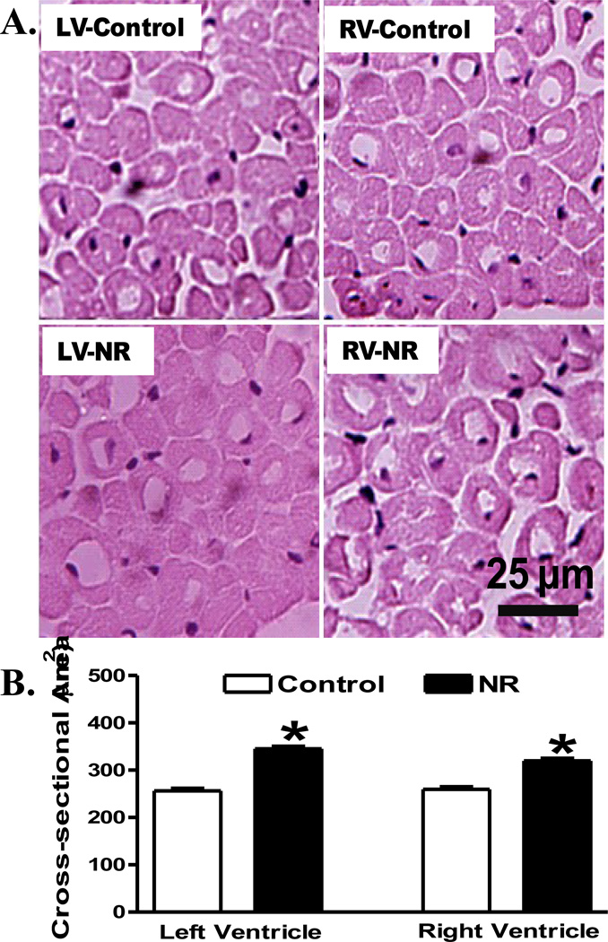
Fig 2.
Histological examination of left ventricle (LV) or right ventricle (RV) from adult offspring of control and nutrient restricted (NR) ewes using hematoxylin and eosin (H&E) staining. A: Representative H&E staining images from LV and RV in control and NR groups; B: Quantitative analysis of cardiomyocyte cross-sectional area. Averaged areas of at least 200 nucleated myocytes per section were used from each sheep. Mean ± SEM, n = 4; * p < 0.05 vs. respective control group.

