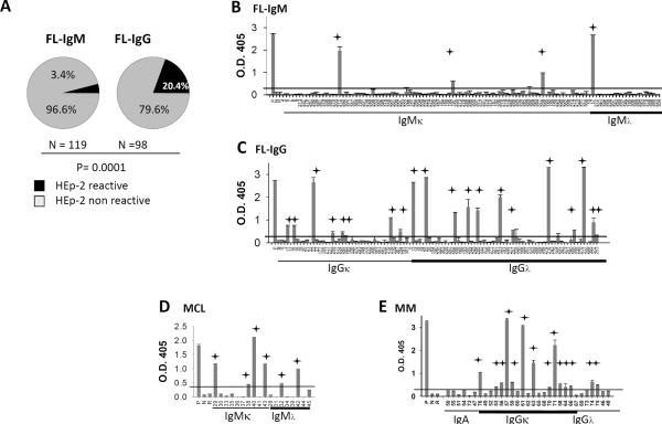
Figure 2. Tumor Igs are frequently self-reactive.
FL Igs were tested in triplicate for reactivity against HEp-2 whole-cell lysates using ELISA as described in Materials and Methods. (A) The percentage of IgM+ and IgG+ FL Igs reacting to HEp-2 whole-cell lysates. P was calculated using a 2 × 2 Fisher's exact test. (B–E) Reactivity of individual tumor Igs from (B) IgM+ FL (n = 119), (C) IgG+ FL (n = 98), (D) MCL (n = 18), and (E) MM (n = 31) against HEp-2 whole-cell lysates is shown by optical density (O.D.) determined at 405 nm. Each tumor Ig is indicated by a unique number, and a polyreactive ED38 antibody was used as a positive control (P). Nonreactive mG053 antibody (N), and rituximab (R) were used as negative controls. Tumor Igs were considered to be HEp-2-reactive if the O.D. was greater than 5 times the O.D. of the nonreactive mG053 antibody (indicated by red line). Error bars represent the standard deviation for triplicate samples, and the stars indicate HEp-2-reactive tumor Igs. Data were reproducible in three independent experiments.

