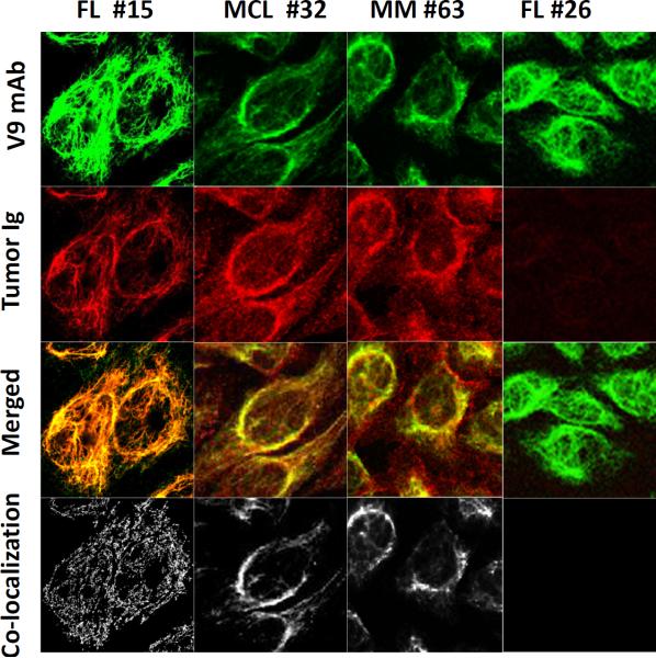
Figure 4. Vimentin-reactive tumor Igs colocalize with anti-vimentin V9 mAb.
Immunofluorescence staining of HEp-2 cells was performed with tumor Igs from FL15, FL26, MCL32, and MM63, and colocalization with anti-vimentin V9 mAb was determined. Each column represents staining with one tumor Ig. Top to bottom: V9 mAb alone (green), tumor Ig (red), V9 mAb and tumor Ig overlay, and colocalization pixels.

