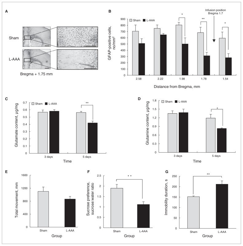
Fig. 1.
L-α aminoadipic acid (L-AAA) infusion into the mouse prelimbic cortex (PLC) led to a reduction of astrocytes compared with the vehicle injection group (sham; A and B, bregma 1.98, t8 = 2.86, p = 0.021; bregma 1.78; t = 3.63, p = 0.007; bregma 1.54, t8 = 2.68, p = 0.028). We performed the immunohistochemical analysis on the sixth day after the first L-AAA infusion. Glutamate (Glu) and glutamine (Gln) levels in the prefrontal cortex (PFC) were measured after L-AAA infusion. There was no change in Glu or Gln levels in the PFC on the third day after the infusion (Glu level of day 3, t8 = −0.840, p = 0.42; Gln level of day 3, t8 = −0.174, p = 0.87); however, Glu and Gln levels in mice infused with L-AAA decreased significantly compared with those of control mice on the fifth day after L-AAA infusion (C and D, Glu level day 5, t8 = 3.488, p = 0.008; Gln level day 5, t8 = 2.596, p = 0.032). L-α aminoadipic acid infusion did not affect the locomotor activity in the open field test (OFT; E, t8 = 1.505, p = 0.17). Sucrose preference from the sucrose preference test (SPT) was significantly decreased (F, t8 = 3.585, p = 0.007) and the duration of immobility from the forced swim test (FST) increased (G, t8 = −3.791, p = 0.005) in mice infused with L-AAA on the fifth day after infusion, indicating a greater degree of depressive behaviour. The OFT and FST were sequentially performed using the same animals each day. Data are presented as means and standard errors of the mean. We used the t test to evaluate the mean difference between the control mice and those infused with L-AAA. *p < 0.05, **p < 0.01, n = 5 per group. Scale bars in A are 500 μm. GFAP = glial fibrillary acidic protein.

