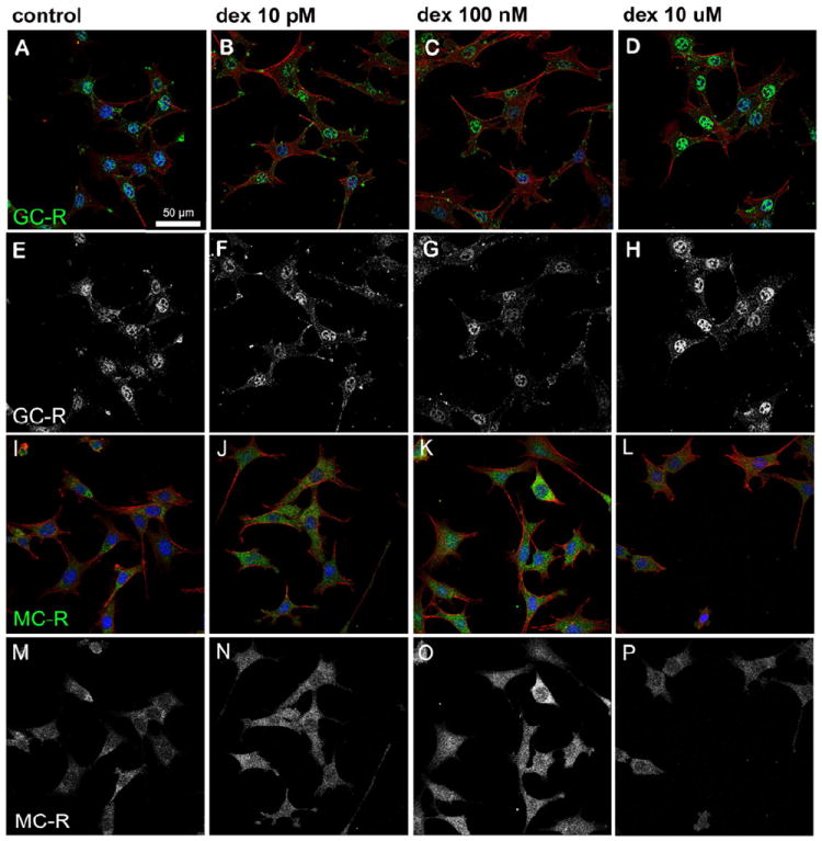
Figure 10. Dexamethasone induced nuclear translocation of GC-R and MC-R in HEI-OC1 cells.

A significant (p≤0.05) increase in nuclear GC-R expression was observed in cells treated with 10 μM dexamethasone (D, H) when compared to untreated cells (A, E) or cells exposed to 10 pM (B, F) or 100 nM (C, G) dexamethasone. A significantly higher (p≤0.0016) nuclear expression of MC-R was observed in cells treated with 100 nM dexamethasone (K, O) when compared to untreated cells (I, M) and cells treated with 10 pM (J, N) or 10 μM (L, P) dexamethasone. Actin is shown in red and nuclei in blue in panels A-D and I-L, whereas GC-R and MC-R are shown in green in panels A-D and I-L, respectively, or white in panels E-H and M-P.
