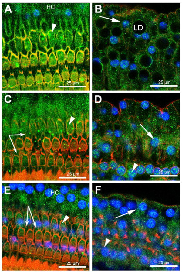
Figure 4. OHCs, Deiters and Hensen cells express GC-R.

Immunofluorescence techniques showed expression of GC-R in the organ of Corti at the apical (A, B), middle (C, D), and basal (E, F) turns of the cochlea. In all these regions GC-R expression was detected in the nucleus and cytoplasm of OHCs, Deiters and Hensen cells. Arrowheads indicate OHCs, arrows Hensen cells, and small arrows Deiters cells. GC-R expression is shown in green, nuclei in blue and actin in red.
