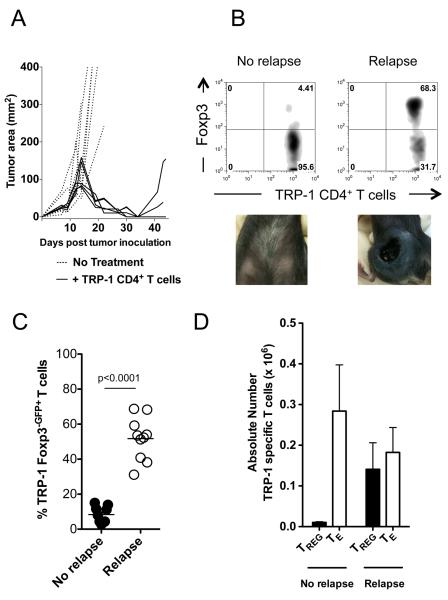
Figure 1. Foxp3+ tumor-specific Treg cells increase during melanoma recurrence.
(A) After successful tumor-specific CD4+ T cell adoptive cell immunotherapy melanoma recurs. C57BL/6 lymphopenic RAG−/− mice (5 mice/group) were inoculated with B16.F10 melanoma (2 × 10⁵ cells). Tumor-bearing mice were treated with 2 × 10⁵ naïve TRP-1 CD4+ T cells by intravenous tail vein injection on day 10 after tumor inoculation. Tumors were followed until recurrence of melanoma. Experiments repeated at least 10 times. (B) Foxp3+ tumor-specific CD4+ T cells increase during relapsing melanoma. TRP-1 CD4+ T cells were analyzed for Foxp3 expression by flow cytometry from relapsing mice (68.3%) and compared to cells from mice with no tumors (4.4%). Data represent ten independent experiments. (C) Percent Foxp3-eGFP+ cells from ten experiments. Using Foxp3-DTR TRP-1 T cells, which fluoresce with eGFP when expressing Foxp3, similar results were obtained in relapsing and non-relapsing mice (No recurrence, ~5%; Recurrence ~50%). P < 0.0001 for Foxp3 expression between non-relapsing versus relapsing mice. (D) Absolute numbers of tumor-specific CD4+ Treg and Foxp3− CD4+ TE cells during recurrence compared to nonrelapsing mice. Experiments repeated ten times.

