
Figure 6.
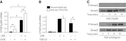
Mesothelial CTGF expression induced by LPA-LPA1 signaling is independent of de novo protein synthesis, latent TGF-β activation, and Smad signaling. A) Effect of cycloheximide (CHX) on LPA-induced CTGF expression. PMCs were preincubated with CHX (20 μg/ml) or control medium for 2 h, and then stimulated with control medium or LPA (10 μM) for an additional 2 h (n=3 cell preparations/group). B) Effect of TGF-β neutralization on LPA-induced CTGF expression. PMCs were exposed to LPA (10 μM, for 2 h) or TGF-β1 (1 ng/ml, for 4 h), with or without a pan-specific TGF-β neutralizing antibody (Ab, 10 μg/ml; n=3 cell preparations/group). C) Ability of LPA vs. TGF-β to induce Smad phosphorylation. PMCs were incubated with LPA (10 μM) or TGF-β1 (5 ng/ml), and their lysates were assayed for phosphorylated Smad3 by Western blot. The experiment was performed in 2 independent series of PMC preparations. Data are expressed as mean ± sem copies of CTGF mRNA relative to copies of β2 microglobulin mRNA. *P < 0.01, **P < 0.05.