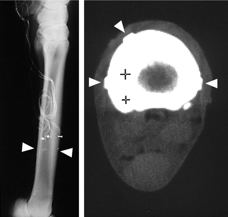
Figure 1.
Left panel: Radiograph of the horse MCIII, showing the level of attachment of the three 3-element rosette strain gauges (arrow showing plane of three gauges, evident by small solder dots visible as small white dots). Approximately 2.5 cm proximal to the gauge site, a plastic flange is used relieve strain on the wires, anchored to the bone via a 2.0-mm screw tapped into the cortex. These radiographs also determine the orientation of the gauge placement (visible at higher magnification) relative to the longitudinal axis of the bone. Right panel: areal properties of the bone, as well as the relative location of the 3 gauges around the cortex (rectangular gauge backing shown by arrows), is determined by CAT scan, performed immediately following surgery while the animal remains anesthetized. The soft tissue (e.g., muscle, skin, and tendons) can be seen as an elliptical silhouette surrounding the bone. Posterior (ventral) surface of the MCIII is toward the bottom, and the medial aspect is to the left. The + symbols on the scan are provided by the computed topography instrument to establish a scale of 1 cm.

