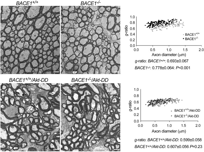
Figure 3.
Myelin sheath was thickened in BACE1-null mice if Akt-DD was overexpressed. Fixed optic nerves were examined by electron microscopy; a representative electrograph from each genotype of mice is shown. Visibly, mice expressing Akt-DD have a much thicker myelin sheath and hypomyelination in BACE1-null optic nerves was clearly reversed. Relative thickness of the myelin sheath can be compared based on the g-ratio calculation. A scatter plot of the g ratios of myelinated axons shows quantitative evidence of hypomyelination in the optic nerves of BACE1–null mice compared with wild-type controls. The g ratio in BACE1−/−/Akt-DD was similar to that in BACE1+/+/Akt-DD (n=3 animals, P=0.23). Scale bars = 1 μm.

