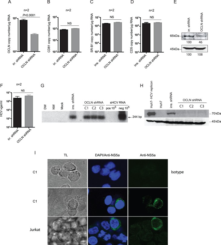
Figure 5. Effect of OCLN knockout on infection of Jurkat T cells with native, patient-derived HCV.
Jurkat cells transduced with either OCLN-specific shRNA or control irrelevant (irr) shRNA were infected with plasma-derived HCV, as described in Materials and Methods. The levels of transcription of (A) OCLN, (B) CD81, (C) SR-B1 and (D) CD5 were evaluated at day 7 post-infection using appropriate real-time RT-PCR assays. The data are presented as mean copy numbers±SEM/µg total RNA from 2 independent experiments each evaluated in triplicate. (E) Western blot detection of OCLN protein in Jurkat cells transduced with either irr-shRNA or OCLN shRNA. Detection of β–actin served as a loading control. The level of OCLN protein in the cells transduced with OCLN shRNA is presented as a relative percentage of the OCLN protein signal detected in control cells that was taken as 100%. (F) Quantification of HCV RNA positive strand in Jurkat T cells transduced with either control irr-shRNA or three separate clones of OCLN shRNA and subsequently infected with wild-type HCV, as described in Materials and Methods. The data represent the mean copy (vge) numbers±SEM from 2 independent experiments each evaluated in triplicate. (G) Detection of HCV RNA replicative strand in Jurkat T cells transduced with either irr-shRNA or three different clones of OCLN shRNA (C1–C3) and infected with HCV, as indicated in (F). Synthetic HCV RNA positive (pos) and negative (neg) strands at 105 copies/reaction served as the assay specific controls. DW, NW and a mock as described in the legend to Figure 1. (H) Detection of HCV E2 protein by Western blotting in Jurkat T cells transduced with irr-shRNA or with three OCLN shRNA clones (C1–C3) and then infected with HCV. Huh7 cells expressing HCV AB12-A2FL replicon and naïve Huh7 cells served as HCV E2 positive and negative controls, respectively. Detection of β-actin served as a loading control. (I) Detection of HCV NS5a protein by confocal microscopy in HCV infected Jurkat transduced with C1encoding OCLN-specific shRNA (C1) and in intact (untransduced) Jurkat cells infected with the same virus as a positive staining control. HCV-infected Jurkat transduced with C1 clone exposed to isotype antibody control instead of anti-NS5a served as a negative control. The cells were counterstained with DAPI to identify nuclei and examined under transmitted light (TL). The images were captured at X60 magnification.

