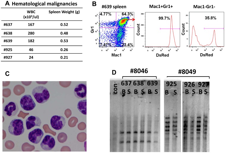
Figure 4. Characterization of myeloid malignancies seen in the SFFV-treated secondary recipients.
(A) Hematologic characteristics of myeloid leukemias including peripheral leukocyte counts and spleen weights in individual mice at the time of euthanasia. (B)Flow cytometry analysis of splenic tumor cells from mouse #639. Staining for the Gr1 and Mac1 myeloid markers is shown. Gated normal cells (Gr1− Mac1− ) and tumor cells (Gr1+ Mac1+) were also analyzed for expression of the DsRed, vector-encoded marker. (C) Peripheral blood smear from a leukemic mouse with abnormal monocytic and granulocytic cells. (D) Southern blot analysis of DNA from bone marrow (B) and spleen (S) of six secondary mice that were derived from two primary recipients is shown. A clonal analysis for vector insertion sites was performed using a single cutter enzyme (BglII) and probed with DsRed cDNA probe. A common clonal pattern was noted from tumors derived from each primary recipient. All mice but #926 had clinical evidence of leukemia, however it was noted that #926 had already converted to a clonal pattern.

