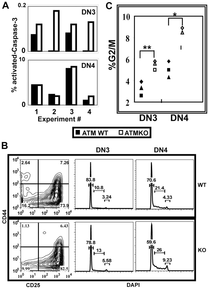
Figure 4. ATM deficiency alters cell survival and proliferation in DN cells.
Freshly explanted thymocytes were stained for surface molecules, fixed, and permeabilized prior to intracellular staining. (A) As compared to ATMWT, ATMKO DN3 but not DN4 cells have an increased frequency of cleaved-Caspase-3+ cells (ATMWT vs KO DN3 p<0.03). Graphs show frequencies of ATMWT (grey bar) and KO (white bar) lineage negative DN3 and DN4 cells that express cleaved-Caspase-3. Data from four independent experiments analyzing ATMWT and KO pairs is shown. (B) Panels show representative lineage negative CD25 CD44 DN staining profiles for ATMWT (top panel) and KO (lower panel) thymi and DAPI staining profiles gated on DN3 and DN4 cells. (C) Frequency of DN3 and DN4 cells from individual ATMWT (black symbols) and KO (open symbols) mice that are in G2/M of the cell cycle. ATM deficiency results in increased cycling cells in both DN3 and DN4 stages (p<0.002 and 0.02, respectively). P values were calculated using Student’s paired 1-tailed t test from data collected from three-four ATMWT and KO pairs analyzed in three independent experiments.

