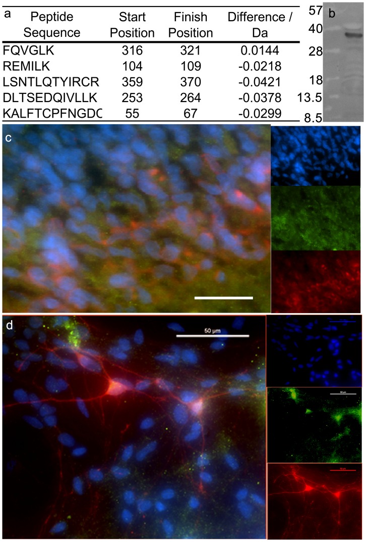
Figure 1. Expression on vitamin D receptor protein.
Vitamin D receptor protein was identified in developing ventral midbrain tissue harvested from embryonic rats. A) Five unique peptides were identified and used to identify VDR. B) VDR protein was also identified by a single band in Western blots of whole tissue lysate obtained from E12 VM of rats. C) Immunohistochemistry of E13 sagittal sections taken through the midbrain show overlapping expression of vitamin D receptor (VDR, green) and tyrosine hydroxylase (TH, red). Scale bar: 20 µM. D) Co-expression of VDR and TH was also observed in single cell cultures of E12 VM tissue. Total cells were labeled with DAPI (blue). Scale bar: 50 µM.

