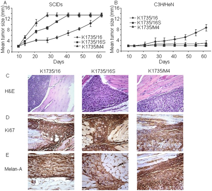
Figure 3. Tumorigenic potential and immunohistological characterization of murine melanoma cell lines in SCID and syngeneic mice using intra footpad injections.
(A) Melanoma cell lines (2×10⁵) injected to the footpad of SCID mice. The K1735/M4 cell line showed the highest tumor growth kinetics, followed by the K1735/16S cell line. The K1735/16 cell line had the slowest tumor growth (n = 5–6 per group, P<0.001). (B) The same concentration of cells injected into the footpad of syngeneic C3H/HeN mice. This time, the K1735/M4 and K1735/16S showed minimal tumorigenic potential, whereas the K1735/16S cell line had the highest tumor growth kinetc (experiments performed 3 consecutive times with n = 10–15 animals per group; P<0.001 between the K1735/16 and the K1735/M4 or the K1735/16S cell lines). (C) Representative histologic features of tumors grown in immune-competent animals, 16 days after implantation. (D) Immunohistochemical analysis with anti-Ki-67 Ab (a proliferation marker) showing similar expression in the three cell lines. (E) Immunohistochemical staining with anti-Melan A Ab (a melanoma marker) showed positive expression in all tumors but not in adjacent normal tissue.

