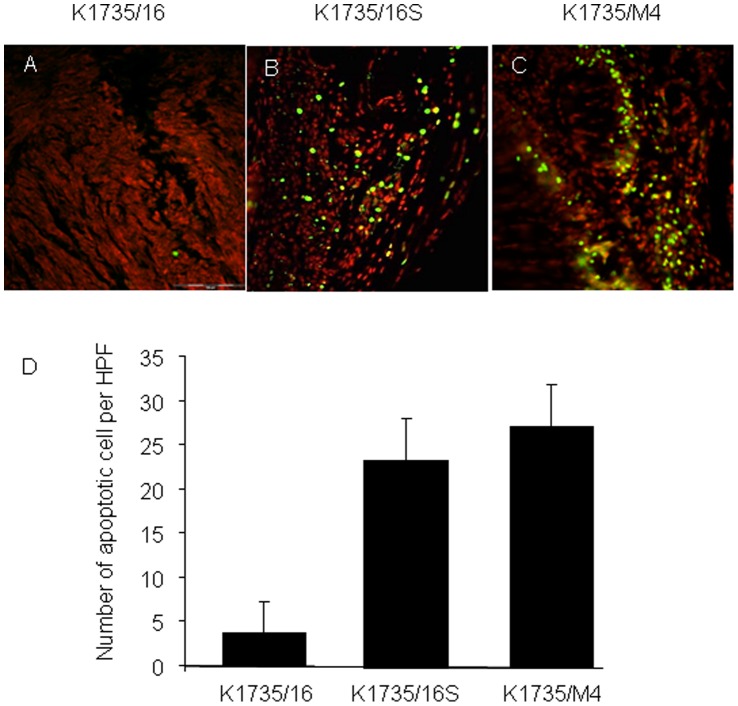
Figure 5. Apoptosis rate of murine melanoma cells in a syngeneic intra footpad model.
Apoptosis was assessed using the TUNEL assay. (A–C) Representative microscopic pictures (magnification X20) of FITC-positive apoptotic tumor cells (green) and counter staining with propidium iodide (red). (D) Percentage of apoptotic cells was quantified by the mean number of FITC-positive cells in 6 microscopic high power fields. Data are expressed as the mean+SD (n = 6 tumors, P<0.001). TUNEL analysis revealed that the number of apoptotic cells in K1735/16 cell line-derived tumors was significantly lower than in K1735/16S and K1735/M4 cells lines-derived tumors.

