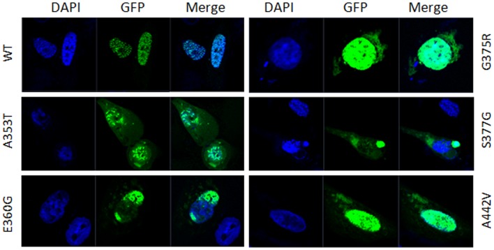
Figure 2. Subcellular location of GATA4 wild type and mutant proteins.
GFP (green) represents the over expressed GATA4 protein, and DAPI (blue) represents the location of the nucleus. The subcellular localization of pEGFP-GATA4 wild-type (WT) and mutant-type proteins shows that wild-type GATA4 completely localized to the nucleus, while mutated GATA4 proteins were partially distributed in the cytoplasm besides the nucleus.

