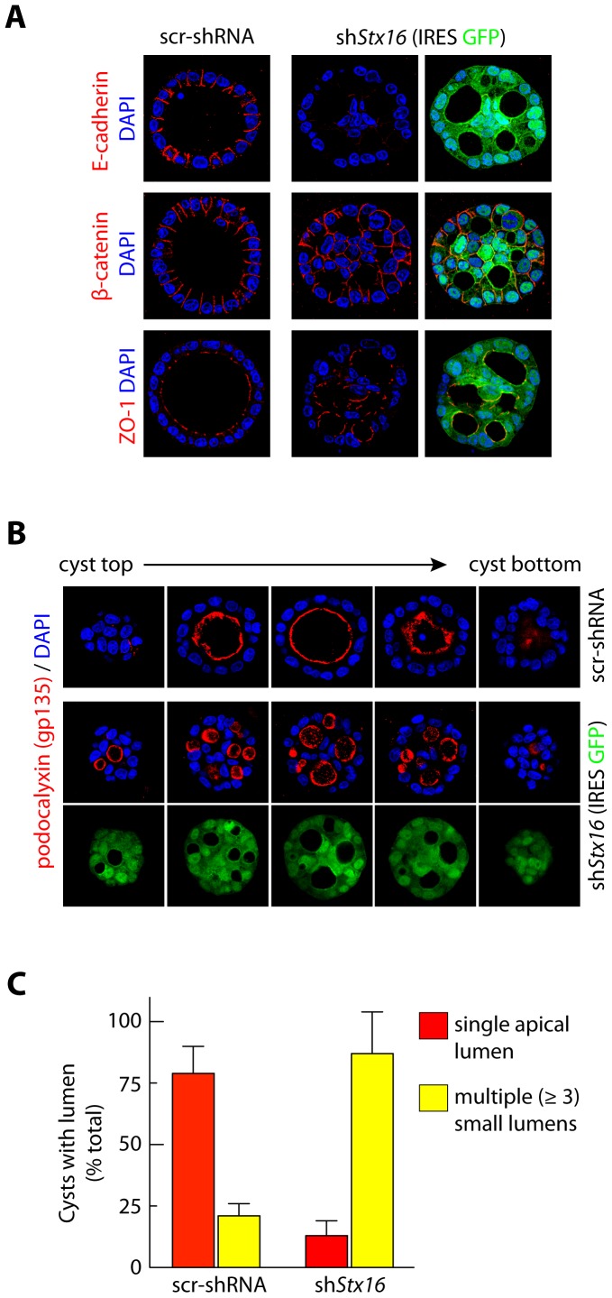
Figure 5. Stx16 depletion leads to decrease in E-cadherin levels and formation of multiple lumens in epithelial cysts.
(A, B) Immunoflourescence-based analysis of morphology and localization of basolateral and apical proteins in cysts formed from MDCK cells stably expressing scr-shRNA or shStx16 4d after plating in Matrigel. Cysts were fixed and immunostained for E-cadherin, β-catenin, ZO-1, and podocalyxin (gp135), and DNA was visualized using DAPI. Scale bar, 5 µm. Representative cysts; confocal sections through the top to bottom of a cyst are shown. (C) Quantification of cysts with single or multiple lumens as in A, B. Values are mean ± SD from three independent experiments; n >100 cysts/experiment. p<0.01.

