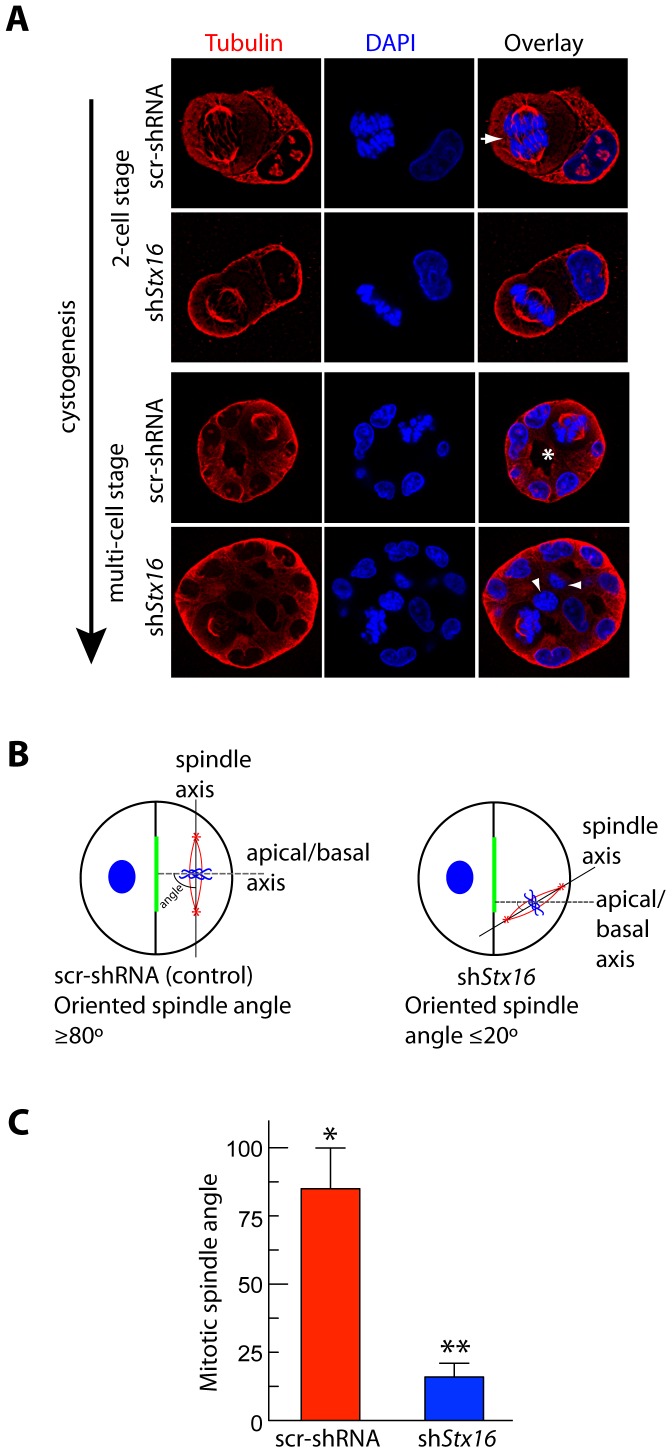
Figure 6. Stx16 depletion leads to spindle misorientation and cystogenesis defects.
(A) Confocal images of metaphase cells in the middle region of the cysts grown for 1–4 d, following fixation and staining with anti-α-tubulin antibody (red) and DAPI (blue). Note that in control cysts: orientation of the spindle with respect to the apical-basal axis is perpendicular (arrow);cell division occurs in the plane of the monolayer (2-cell stage); abscission is asymmetrical; and deposition of nascent apical surface occurs around a single central lumen (asterisk, multi-cell stage). (B) Schematic illustration of spindle-angle measurement. A line was drawn to connect the two spindle poles (thick black line). Another was drawn from the centroid of the apical domain to the midpoint of the spindle axis (thin black lines), and the acute angle (red) between the two lines was assessed. (C) Quantification of metaphase spindle angles in MDCK cells stably expressing scr-shRNA or shStx16. Data show means ± SEM, n = 70 cysts from three independent experiments. Statistical significance was evaluated using a Mann-Whitney test. *, p≤0.002; **, p≤0.001.

