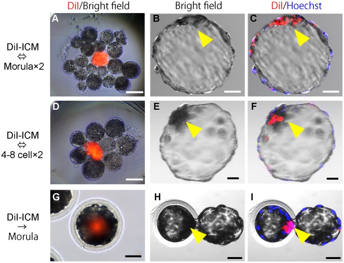
Figure 2. Production of chimeric blastocysts with donor ICM and parthenogenetic host embryos.
(A, D) A donor ICM (stained with Dil) aggregated with host blastomeres isolated from parthenogenetic embryos at the morula (A) or 4–8 cell stage (D). (B, E) Bright field images of chimeric blastocysts developed from the aggregated embryos. (C, F) Confocal fluorescence images of chimeric blastocysts showing DiI fluorescence in ICMs. Single confocal sections of fluorescence were overlaid on the bright field images. (G-I) Parthenogenetic host morulae injected with DiI-stained donor ICM (G) and resultant chimeric blastocysts (H, I). Arrow heads, ICM. Scale bars = 50 µm.

