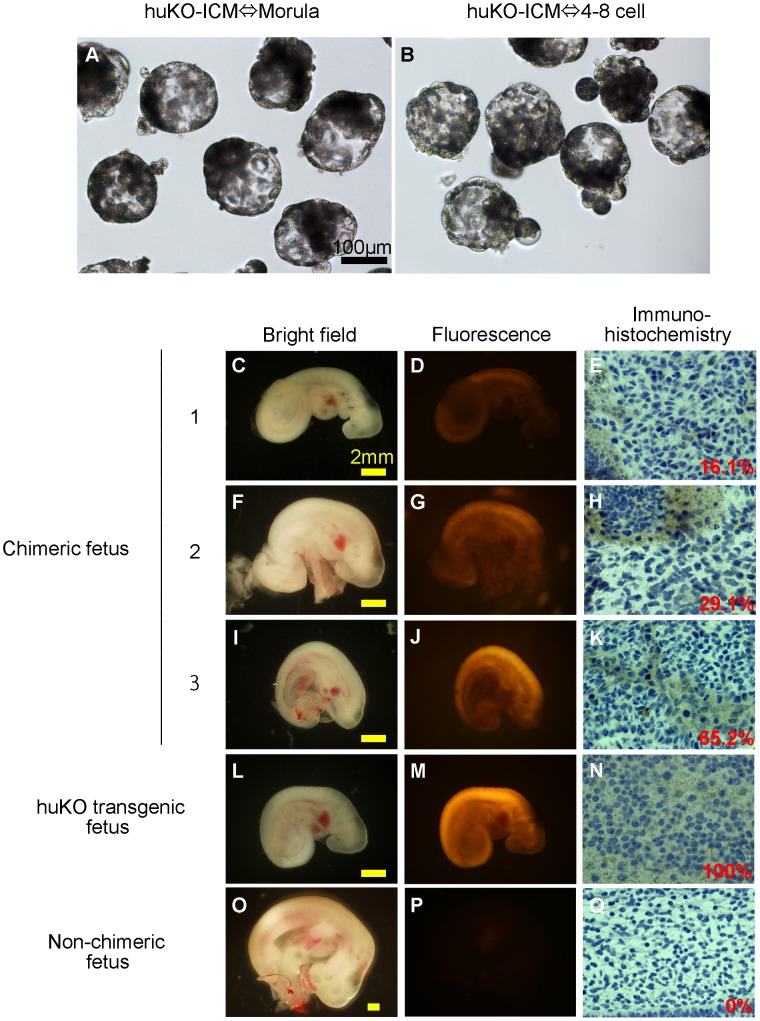
Figure 4. Chimeric fetuses produced by aggregation of the ICM carrying huKO transgene and parthenogenetic host embryos.
(A, B) Morphological appearance of the chimeric blastocysts before embryo transfer. (C–K) Chimeric fetuses (day 18) showing huKO fluorescence derived from the donor ICM cells (C, D, F, G, I, J) and immunohistochemical images showing proportion of the donor-derived (huKO-positive) cells in the tissue of chimeric fetuses (E, H, K). (L, M, N) A day-19 fetus developed from an embryo fertilized in vitro with the huKO transgenic boar sperm as a positive control, showing the systemic expression of huKO (M, N). (O, P, Q) A non-chimeric fetus (day 22) developed from the aggregates of two parthenogenetic embryos as a negative control.

