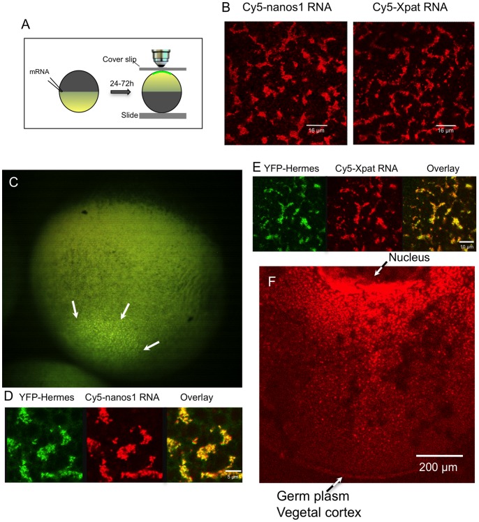
Figure 1. Localisation of ‘early pathway’ mRNAs and Hermes protein into the germ plasm of stage VI oocytes.
A. The procedure for examining the cortical distribution of labelled RNAs. After injection and incubation of oocytes in OCM (with or without vitellogenin-containing serum) for 24 to 72 h, oocytes were held in an inverted position between a slide and a coverslip, in a chamber made with a latex spacer. They were then examined by confocal microscopy using a 40× oil-immersion lens. B. Full-length Cy5-labelled nanos1 and Xpat RNAs localise in islands of particles at the vegetal pole 48 h after injection. C. Low power stereo microscope view from the side of the vegetal pole of a whole stage VI oocyte, 24 h after injection with mRNA encoding YFP-Hermes. YFP-Hermes protein is clearly localised to a field of fluorescent islands at the vegetal pole (white arrows), typical of germ plasm markers. In the stereo microscope the depth of focus is large so that a much larger area than that occupied by the germ plasm is in focus. D,E. In these islands Cy5-labelled nanos-1 and Xpat RNA’s co-localise with YFP-Hermes protein, following co-injection of mRNA encoding YFP-Hermes. All the above oocytes were visualized live, as they are in later figures unless otherwise stated. F. Internal distribution of Cy5-nanos1 RNA between the nucleus and the vegetal cortex of a fixed stage VI oocyte. Following injection of RNA, oocytes were cultured for 48 h in OCM, fixed and hemisected with a scalpel prior to visualization by confocal microscopy using a Z-stack.

