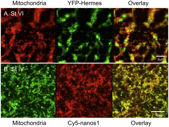
Figure 2. YFP-Hermes localises into particles in islands containing concentrated mitochondria.
A. Stage VI oocytes were injected with RNA encoding YFP-Hermes and after 18 h mitochondria were stained with TMRE [22], prior to visualisation of the vegetal cortex, as in Figure 1A. B Stage IV oocytes were injected with Cy5-nanos1 RNA and after 24 h the oocytes were stained with TMRE and analysed as in A.

