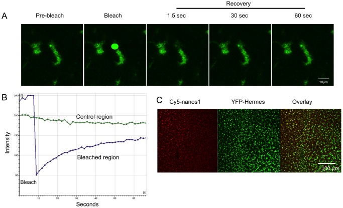
Figure 5. FRAP experiment showing that Hermes protein in germ plasm exchanges with a cytoplasmic pool YFP-Hermes.
A. Confocal images, before during and after the bleaching step. B. Quantification of the bleached area and a small unbleached control region. C. Low power image to show that injected RNA diffuses more slowly than protein. Oocytes were co-injected at the equator with Cy5-nanos1 and YFP-Hermes RNAs (The latter does not have its own UTRs, so would not localise). After 48 h the vegetal pole was visualised and it is seen that the Cy5-nanos1 RNA has diffused more slowly from the injection point, at the top left, than Hermes protein.

