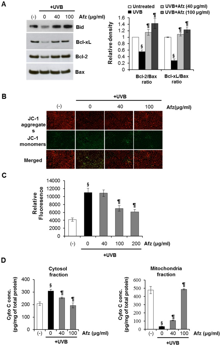
Figure 6. Afzelin inhibits UVB-induced mitochondrial effects.
A. Effect on UVB mediated changes on bid, Bcl-xL, Bcl-2, and Bax protein expression. HaCaT cells were irradiated with UVB (20 mJ/cm2) and incubated for 12 h. Cell lysates were subjected to immunoblot analysis with antibodies against bid, Bcl-xL, and Bax. The Bcl-xL/Bax and Bcl-2/Bax ratios are presented in graphs. B. Mitochondrial membrane potential (Δψm) was measured by staining HaCaT cells with JC-1 followed by fluorescence microscopy analysis. HaCaT cells were incubated with the JC-1 probe for 30 min. Mean (red/green) fluorescence, expressed as a percentage of the control, indicates the ratio of high/low mitochondrial membrane potential. Representative data, n = 3 (A–B). C. DHR 123 was employed to detect mitochondrial hydrogen peroxide. Reactive oxygen species (ROS)-induced DHR 123 fluorescence was measured using a spectrophotometer. D. Enzyme-linked immunosorbent assay (ELISA) analysis of cytochrome c in cytosolic and mitochondrial fractions of HaCaT cells exposed to UVB (20 mJ/cm2). HaCaT cells were treated with afzelin (40–100 µg/ml) for 12 h after exposure to UVB (20 mJ/cm2) radiation. Then, the cells were processed for cytochrome c analysis. Data are means ± SD, n = 3 (A–D). § P<0.01 compared with the vehicle-treated group, ¶ P<0.01 compared with the UVB -treated group (A–D).

