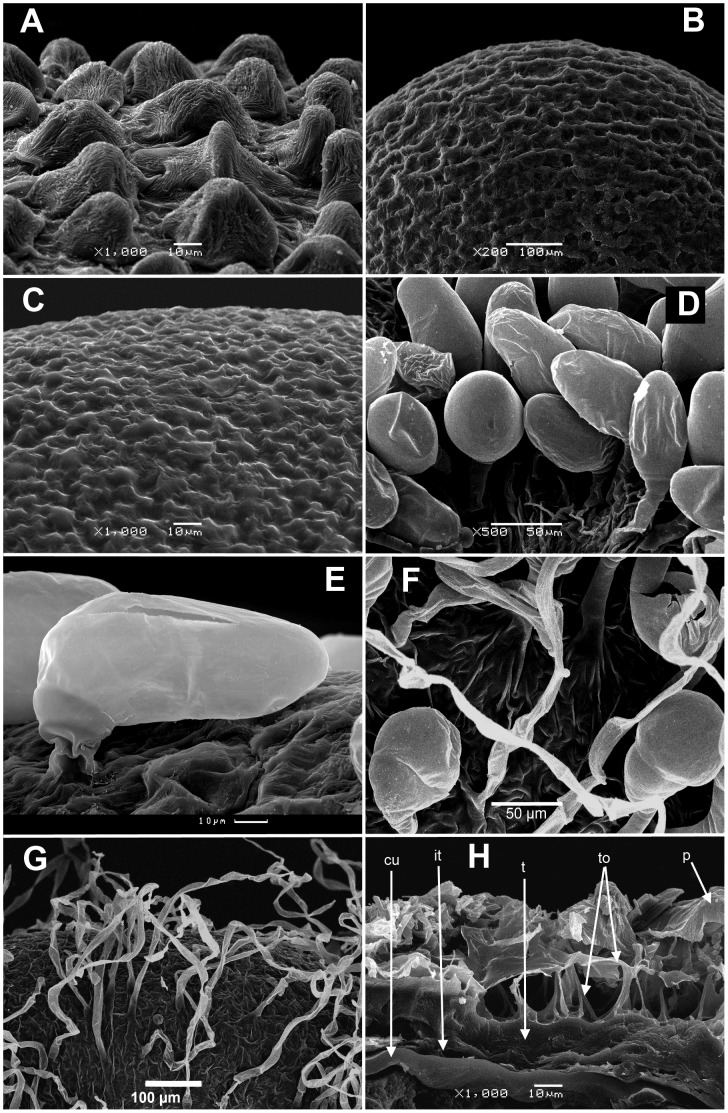
Figure 3. Scan micrographs of the fruits.
(A) Papillae on pericarp surface of Dysphania procera (1000x); (B) Pericarp surface of D. congolana (200x); (C) Tiny papillae on pericarp surface of D. pseudomultiflora (1000x); (D) Glandular trichomes on the pericarp surface of D. chilensis (500x); (E) Glandular trichomes on the pericarp surface of D. multifida (1000x); (F) Glandular trichomes and central parts of curved simple hairs on the pericarp surface of Cycloloma atriplicifolium (500x); (G) Simple curved hairs on the pericarp surface of C. atriplicifolium (200x); (H) Cross-section of the pericarp and seed coat of Blitum atriplicinum (1000x) showing acicular outhgrowths of the testa cells. Abbreviations: p – pericarp; t – testa; to – testa outgrowths; it – integumental tapetum; cu – cuticle between integumental tapetum and perisperm.

