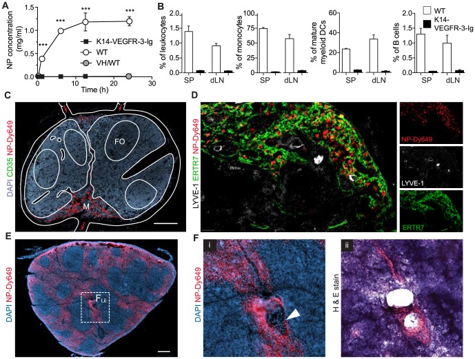
Figure 3. Lymphatic drainage is required for nanoparticle targeting of the lymph node and spleen after i.d. administration.
(a) Bioavailability of Dy649-nanoparticles (NPs) in the blood compartment after i.d. administration in mice that lack peripheral lymphatics (K14-VEGR-3-Ig) and their wild type littermates. VH: vehicle control (non-fluorescently labeled NPs). Two-way repeated measures ANOVA followed by Bonferroni post test. (b) Comparison of NP+ association as assessed by flow cytometry in the brachial lymph node (LN) and the spleen 24 h post-i.d. administration. n = 4 *p≤0.05, ***p≤0.005. (c) 9 µm thick section of a Dy649-NP (red) draining wild type popliteal LN stained with nuclei (DAPI, blue). Scale bar, 200 µm. (d) 9 µm thick section of the NP-Dy649 (red) draining wild type brachial LN 12 h after i.d. administration, stained for lymphatic endothelium (LYVE-1, green), the T cell zone stroma (ERTR7, white). Scale bar, 40 µm. (e) 40 µm section of the wild type anterior spleen stained with DAPI (blue) and NP-Dy649 (red) shows NP accumulation in the red pulp (RP) and the marginal zone (MZ), as well as surrounding the B cell follicles (FO). Scale bar, 300 µm. (f) Enlarged region of the central arteriole of the spleen (white filled arrow) (i) immunofluorescence image (NP-Dy649, red and DAPI, blue). (ii) Hematoxylin & eosin staining of the same section of the spleen; dark blue FO, purple red pulp and pink blood vessels. Scale bar, 100 µm.

