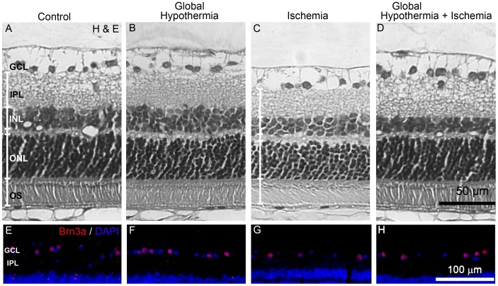
Figure 3. Effect of global HPC on retinal histology.
Upper panel: Representative photomicrographs showing histological appearance of non-ischemic retinas without (A) or with global HPC (B), ischemic retinas 14 days after 40 min of ischemia without global HPC (C), and ischemic retinas from animals submitted to global HPC, 24 h before ischemia (D). Severe retinal damage is shown in the retina from eyes submitted to ischemia without global HPC, whereas in animals submitted to global HPC, the retinal structure was notably preserved. Lower panel: Immunohistochemical detection of Brn3a(+) cells in the GCL from all the experimental groups. A decrease in GCL Brn3(+) cell number was observed in ischemic retinas without global HPC (G) as compared with non-ischemic eyes (E), whereas global HPC, which showed no effect in control eyes (F), partly preserved Brn3a(+) cell count in ischemic eyes (H). GCL, ganglion cell layer; IPL, inner plexiform layer; INL, inner nuclear layer; ONL, outer nuclear layer, OS, outer segment of photoreceptors.

