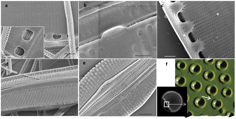
Figure 6. Localization of the organic matrix in N. curvilineta (a, c and d), A. salina (b and e) and C. radiatus (f).
a, b, d and e: SEM of SDS-cleaned valves. a and b: SEM of valve proximal surface showing the organic matrix. d and e: SEM of distal surface showing naked silica. c: SEM of acid cleaned valve proximal surface (compare with a). Scale bar: a, c = 1 µm, d, e = 2 µm, a inset, b = 500 nm. f and inset; AFM and fluorescent micrographs of DAPI-stained SDS-cleaned C. radiatus valve showing the area where the organic layer peeled off (arrow in f).

