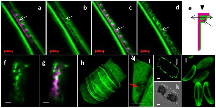
Figure 8. a–g; Fluorescent micrographs of whole cells stained with calcofluor (green) and HCK123 (pink).
a–d; Two different optical sections (a, b and c, d) of dividing N. curvilineata in longitudinal section (scale bars 2 µm). e; sketch of N. curvilineata valve in transversal section showing the orientation of the section (double arrow) and the view (arrow head at top) of a–d. White arrows in a-d show a fibulae surrounded by calcofluor stained material, and correspond to the dark single-headed arrow in e. f and g; Longitudinal section of dividing C. cryptica (scale bars 2 µm). h–k; T. dubium stained with calcofluor (scale bars 5 µm). h; reconstructed 3D image of whole cell. i; Zoom on the matrix associated with the valve showing the absence of matrix in the area corresponding to the ocelli (white arrow) and the spine (red arrow). j and k; optical section through a dividing cell, calcofluor (j) and corresponding DIC (k). l; A. salina stained with calcofluor (scale bar 5 µm).

