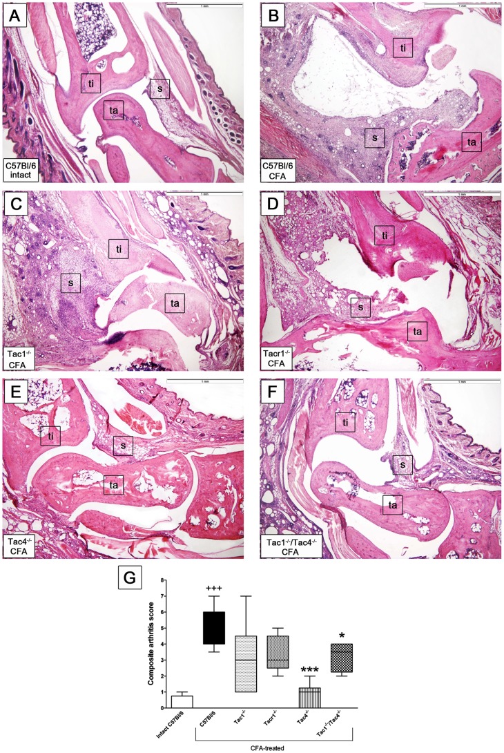
Figure 3. Histopathological changes of the paws on day 21.
Panel A shows a representative histopathological picture of an intact tibiotarsal joint and panel B demonstrates the joint structure of an adjuvant-treated C57Bl/6 wildtype mouse with remarkable synovial swelling, leukocyte infiltration, cartilage damage, and bone destruction. The lower panes demonstrate the joint structures of adjuvant-treated (C) Tac1−/−, (D) Tacr1−/−, (E) Tac4−/−, and (F) Tac1−/−/Tac4−/− mice, decreased inflammatory parameters can be observed in the latter two groups. Hematoxylin-eosin staining, 40x magnification (ti: tibia, ta: tarsus, s: synovium). (G) Semiquantitative histopathological scoring on the basis of inflammatory cell accumulation, synovial enlargement, cartilage destruction and bone erosion. Box plots represent the composite scores (n = 4–12 mice per group,+++p<0.001 vs. intact C57Bl/6, *p<0.05, ***p<0.001 vs. C57Bl/6 CFA-treated, Kruskal-Wallis followed by Dunn’s post test).

