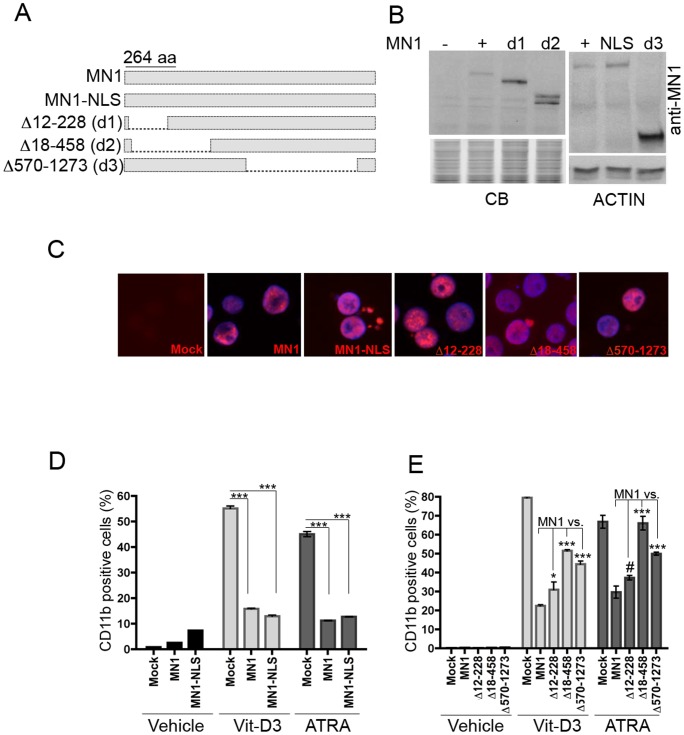
Figure 1. Effects of MN1-deletion mutants on myeloid differentiation of U937 cells.
(A) Schematic representation of MN1 and MN1-deletion mutant proteins. (B) Western blot analysis of GFP-sorted U937 stable cell lines overexpressing the indicated proteins. Figure shows immunoblotting results of anti-MN1 antibodies recognizing either the N-terminus (top left panel) or C-terminus of the protein (top right panel) and anti-human ACTIN antibody (bottom right panel). The commassie-blue stained image of the PAGE gel (bottom left panel; CB) and a pan-actin blot (bottom right panel; ACTIN) are shown as loading controls. (C) Immuno-cytochemical analysis of the above cells using the same MN1-antibodies (red). Blue shows nuclear staining with 4,6-diamidino-2-phenylindole (DAPI). Images were captured with an Olympus BX-50 microscope (equipped with UPlanFL 40×/0.75 numerical apertures with a SPOT camera and SPOT Advanced Imaging software of Diagnostic Instruments. The original magnification was x400. (D, E) FACS analysis showing CD11b expression of indicated U937 stable cell lines 3 days after treatment with vehicle, vitamin D3 (Vit-D3) or ATRA (mean ± SEM of duplicates; *P<.05, ***P<.001, #not significant).

