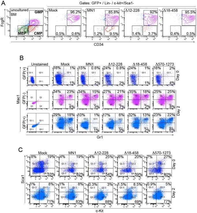
Figure 2. Deletion of aa residues between 18–458 or 570–1273 of MN1 restores MN1-impaired myeloid differentiation of mouse HSPC in vitro.
(A) FACS analysis showing the distribution of MEP, CMP and GMP populations in lineage depleted mouse bone marrow HSPC that were transduced with retroviruses expressing GFP alone (mock), MN1, or the indicated MN1-deletion mutants. Freshly isolated bone marrow cells were used to set up the gates. Figure shows one representative results from 3 independent experiments. (B) Mouse HSPCs transduced with indicated retroviruses were subjected to a second lineage depletion 96 hrs after transduction (Day-0) and cells were cultured for an additional 2 days (Day-2). FACS analysis shows Mac1 and Gr1 expression of unsorted cells at Day-0 and Day-2 of culture. (C) FACS analysis showing the Sca1 and c-Kit expression of the GFP (+) cells in panel B.

