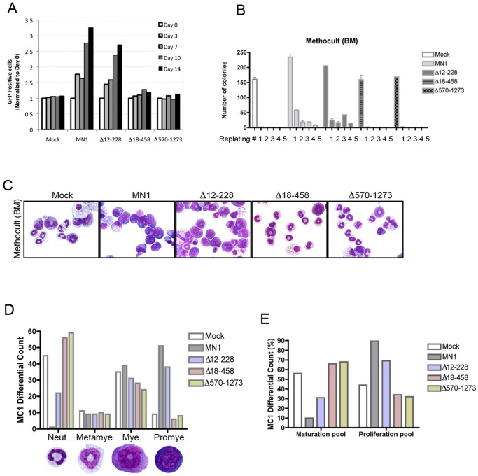
Figure 3. Amino acid residues 12–228 of MN1 are not required for its proliferative and self-renewing activity in vitro.
(A) Periodic GFP analysis was performed at indicated times of liquid cultures using FACS. GFP percentage at Day-0 of analysis (4 days after the last transduction) in each sample was set to 1 and the results of all indicated time points were calculated relative to Day-0. Graph shows one representative analysis of three independent experiments. (B) Methylcellulose assays showing colony numbers after serial replating (1 to 5) of linage-depleted mouse HSPCs transduced with the indicated retroviruses (mean ± SEM of duplicates). (C) May-Grunwald/Giemsa stained images of cytospins of the cells in panel B (after the first methylcellulose culture). (D, E) Differential-count of cytospins is shown in panel C. A total of 100 cells were counted in each sample. The maturation pool includes neutrophils and metamyelocytes whereas the proliferation pool comprises myelocytes and promyelocytes. The images in panels C and D were captured with an Olympus BX41 microscope, equipped with SPOT Insight Color Mosaic 2 MP camera and SPOT imaging software (original magnification x1000).

