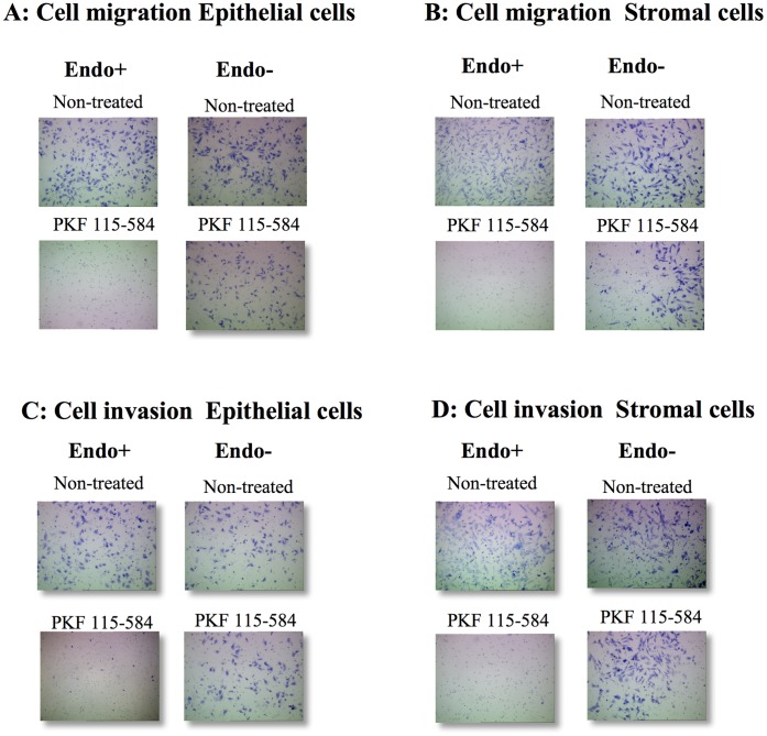
Figure 3. Representative photomicrographs of cell migration and invasion.
A, B: Representative photomicrographs of migration of non-treated and PKF 115–584–treated menstrual endometrial epithelial (A) and stromal (B) cells of patients with and without endometriosis (magnification x100). C, D: Representative photomicrographs of invasion of non-treated and PKF 115–584–treated menstrual endometrial epithelial (C) and stromal (D) cells of patients with and without endometriosis (magnification x100). Endo (+): Endometrium of patients with endometriosis prepared from the menstrual phase. Endo (–): endometrium of patients without endometriosis prepared from the menstrual phase.

