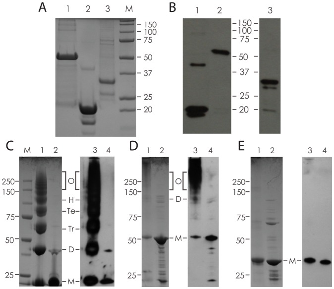
Figure 2. Expression, purification and chemical crosslinking of recombinant proteins.
A) Coomassie Blue staining of purified, His-tagged proteins separated by 12% SDS-PAGE; 1. Acr-Ag85B (50 kDa), 2. Acr (20 kDa) and 3. Ag85B (32 kDa). B) Western blot analysis using antigen-specific antibodies; 1. Acr, 2. Acr-Ag85B and 3, Ag85B (1 and 2 probed with anti-Acr mAb TBG65; 3 probed with rabbit anti-Ag85B serum). C) Chemical crosslinking of Acr; shown is a Coomassie-stained (1, and 2) or Western blot (3 and 4) analysed sample with crosslinker (1 and 3) or without crosslinker (2 and 4). Letters indicate various molecular forms based on expected size (M-monomer, D-dimer, Tr-trimer, Te-tetramer, H-hexamer, O-oligomers). D) Chemical crosslinking of Acr-Ag85B fusion protein; shown is a sample with (1 and 3) or without (2 and 4) crosslinker. Letters indicate various molecular forms as for Acr (C). E) Chemical crosslinking of Ag85B (internal control); Coomassie and Western blot analysis of a sample with (1 and 3) or without (2 and 4) crosslinker.

