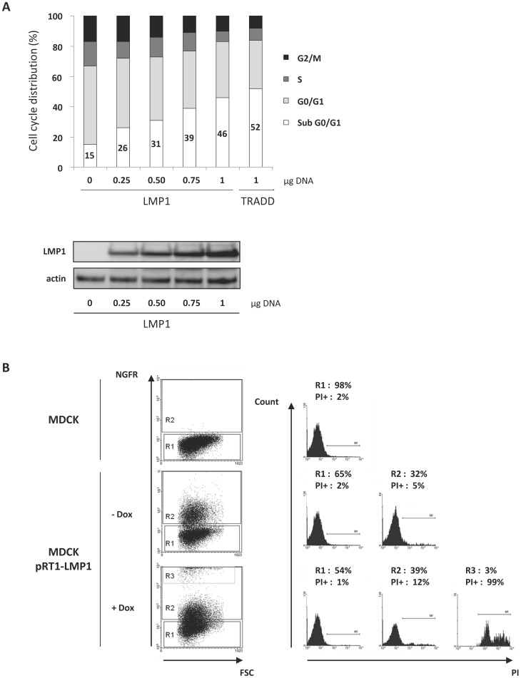
Figure 1. Ectopic expression of LMP1 in MDCK cells induced cell death.
(A) MDCK cells were transfected with increasing amounts (0–1 µg) of LMP1 vector. Cells were then permeabilized with ethanol and labelled with propidium iodide (PI). Cell cycle distribution was analyzed using flow cytometry. TRADD-expressing cells were used as a control of cell death induction. LMP1 expression was analyzed by Western blot. Actin was used as a loading control. (B) MDCK cells were transfected with a doxycyline (Dox)-inducible vector (pRT1-LMP1) endowed with a bidirectional promoter which drives the expression of both LMP1 and a truncated version of NGF receptor (NGFRt) serving as a reporter gene. The detection of NGFRt at the surface of cells implies that these cells also express LMP1. Cells were treated (+Dox) or not (- Dox) with Dox (2 µg/ml) for 24 hours, stained with PI and PE-conjugated antibodies directed against NGFRt, and then analyzed by flow cytometry. Percentages of cells in each cell population (R1: negative NGFRt labelling; R2: low NGFRt labelling; R3: high NGFRt labelling) and corresponding propidium iodide positive cells (PI+) (i.e dead cells) are indicated. Parental MDCK cells were used as a control.

