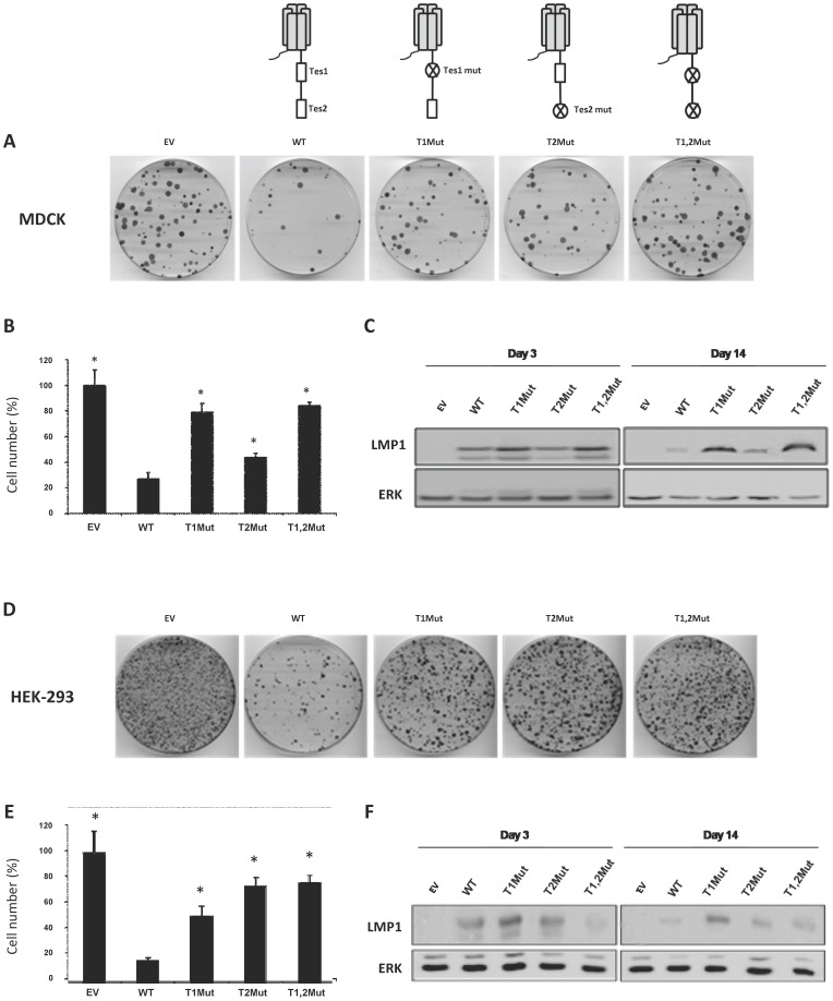
Figure 2. LMP1 induced cell death through its C-terminal tail.
MDCK (A to C) and HEK-293 (D to F) cells were transfected with a empty vector (-) or a vector driving the expression of different versions of LMP1: the wild-type protein (WT) and versions of LMP1 in which either TES1 (T1Mut), or TES2 (T2Mut) or both (T1,2Mut) are altered. Two days later, cells were split into 5 dishes and cultured for 11 days in the presence of G418 to favor the selection of transfected cells. (A, D) Pictures of cells stained with Giemsa. (B, E) Cell counting. Results are represented as the mean of three different dishes, and given as a percentage of cells transfected with the empty plasmid (100%). Standard deviations are shown. *p-value of <0.05, compared to the WT LMP1 expressing vector (WT), two-sample t test (two-tailed). (C, F) Whole cell extracts from cells cultured for 3 days or 11 days in the presence of G418 were prepared. Proteins were resolved by 10% SDS-PAGE and analyzed by Western blot using antibodies directed against LMP1 or ERK2 (used as a loading control).

