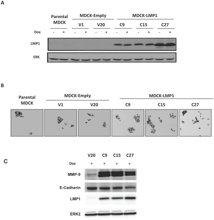
Figure 4. Characterization of MDCK cell lines stably expressing LMP1.
MDCK cells were transfected with the pSTAR encoding or not LMP1 cDNA After selection with G418, two control clones (V1 and V20) and three LMP1-expressing clones (C9, C15 and C27) were selected and further analyzed with (Dox+) or without (Dox-) doxycycline induction. (A) Expression of LMP1 by Western blot analysis. ERK was used as a loading control. (B) Representative pictures of each cell line. Parental MDCK cells are shown as a reference. (C) Expression of LMP1, E-cadherin and MMP9. ERK was used as a loading control.

